Chinese scholars elucidate the mechanism of self-activation and force perception of adhesion-like GPCRs
At 23:00 on April 13, 2022, Beijing time, the team of Professor Jinpeng Sun and Professor Xiao Yu from the School of Basic Medicine of Shandong University published two researches on adhesion G-protein coupled receptors (aGPCR) in the journal Nature at the same time. His work clarified the mechanism of aGPCR self-activation and mechanical force perception, proposed a "finger model" of aGPCR activation, and innovatively conceived a development plan for universal polypeptide ligand antagonists.


G protein-coupled receptors (GPCRs) are the largest family of membrane receptors encoded by the human genome. They are widely distributed in various tissues and organs of the human body and are responsible for nearly half of the transmembrane signal transduction of cells. is the most important drug target. Compared with other members of the GPCR family, aGPCR has remarkable structural features, including a complex multi-domain N-terminal extracellular region (Extracellular region) and GPCR autoproteolysis-inducing (GAIN) that can undergo autohydrolysis. Domain, which contains a conserved GPCR proteolysis site (GPS), most aGPCR members can spontaneously undergo autohydrolysis at the GPS position, resulting in two subunits, α and β, of which the α subunit is the extracellular region (NTF), the β subunit is a region containing seven transmembrane helices (CTF), and the hydrolyzed NTF and CTF remain non-covalently linked on the cell surface. After autohydrolysis of aGPCR, the most N-terminal region of its β subunit is called the Stachel sequence. Studies have shown that aGPCR can sense a variety of external signal stimuli and have distinct activation modes, such as being activated by mechanical forces such as intracellular fluid flow or vibration, and activated by soluble small molecules or protein ligands. The activation of adhesion-like GPCRs caused by Stachel sequences has always been the core content of adhesion-like GPCR signals and functions. How Stachel sequences interact with receptors and the general mechanism for regulating the activation state of receptors is still unclear.
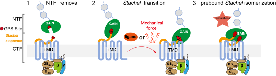
"Force" can determine the fate of cells. The perception of force is very important for living organisms. Mechanical force stimulation drives many physiological processes, which are crucial to the dynamics of cells and the normal growth and operation of the body. In the work of "Structural basis for the tethered peptide activation of adhesion GPCRs", Professor Sun Jinpeng's team used the adhesion receptors GPR133 and GPR114 as models for structural analysis to explore the activation mechanism of aGPCR mediated by Stachel sequence (Figure 1). It was found that GPR133 GPS site undergoes hydrolysis and NTF-CTF separation occurs on the plasma membrane, while GPR114 does not undergo autohydrolysis and can sense mechanical force. And through functional experiments, it was proved that the receptor activates the receptor through the Stachel sequence after sensing the mechanical force. The cryo-electron microscopy structure analysis further confirmed that the conserved HIM (Fss03xφφφxφss09) composed of five hydrophobic amino acids in Stachel plays a role in the interaction between the Stachel sequence and the receptor. It plays a central role in elucidating the structural basis for the recognition of Stachel sequences by receptors, and reveals the activation mechanism of adhesion receptors under the action of mechanical force depending on Stachel sequence activation. In order to gain a deeper understanding, it also provided a basis for the design of exogenous short peptides from Stachel sequences.
In the work of "Tethered peptide activation mechanism of the adhesion GPCR ADGRG2 and ADGRG4", Yu Xiao and Sun Jinpeng's team analyzed the complex structure of Stachel sequence-activated ADGRG2-β-Gs and ADGRG4-β-Gs by cryo-electron microscopy, revealing The direct mechanism of action between Stachel sequence and aGPCR. It was found that the five hydrophobic amino acids of the Stachel sequence were distributed in a finger shape, which played a major role in the activation process mediated by the Stachel sequence. Therefore, a general "finger" model activation mode of aGPCR activation was proposed and named as the finger model (Figure 2). .
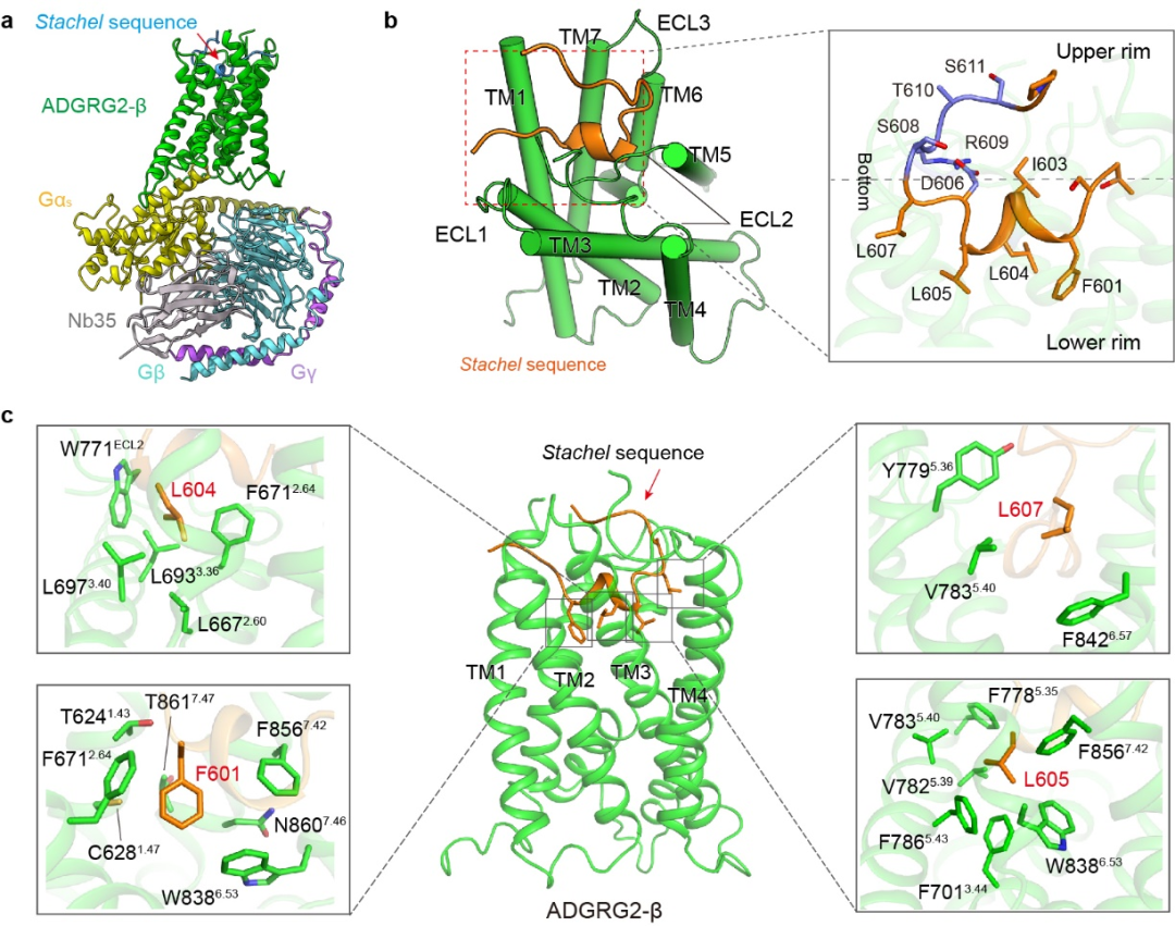
Figure 2: Interaction mode of Stachel sequence with ADGRG2. a. Structural model of the ADGRG2-β-Gs complex activated by the Stachel sequence. b. The Stachel sequence binds to the pocket formed by the seven transmembrane helices of ADGRG2 in a “U” configuration, and is divided into a polar hydrophilic upper half and a hydrophobic lower half along the direction of the bottom opening. c. The five hydrophobic amino acid fingers and positions of the hydrophobic part of the Stachel sequence interact directly with the five hydrophobic pockets of the receptor.
Earlier, the team of Yu Xiao and Sun Jinpeng reported a short peptide agonist ligand VPM-p15 based on the sequence of ADGRG2 Stachel. In this study, combined with the finger model activated by aGPCR, a high-affinity VPM-IP15 ligand was obtained by further optimization and transformation. Based on the cryo-EM structure of IP15 and full-length ADGRG2, the enhanced hydrophobic interaction with the receptor revealed the specific mechanism of the increased affinity between IP15 ligand and full-length ADGRG2. Afterwards, through a series of biochemical experiments, it was found that Fss03 in IP15 was replaced by D/E, making it an antagonist of ADGRG2. This strategy of obtaining polypeptide antagonists by negatively transforming polypeptide agonists, like putting a ring on the index finger of the activated finger model, decisively changed the function of adhesion receptor-like polypeptides (Fig. 3). This method also works for ADGRG4, ADGRG5, and ADGRG6. In this study, Fss03 was modified by polarity, and a general modification method for the antagonistic short peptide ligands of the aGPCR family was developed.
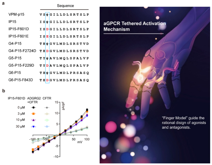
Figure 3: Development of a general peptide antagonist engineering strategy for aGPCRs. a. Sequence alignment of Stachel peptidyl agonists and antagonists. By mutation of Fss03 to Dss03 or Ess03, marked with blue boxes. Ψ: 4-MeF. b. Stachel peptidyl antagonists F601D and F601E affect ADGRG2-mediated CFTR current responses.
The two research works have been guided and strongly supported by Professor Feng Shiqing of Qilu Hospital and Professor Wang Chuanxin of the Second Hospital of Shandong University in terms of subject development, thinking condensed and article writing. Prof. Sun Jinpeng and Prof. Feng Shiqing have carried out a series of collaborations on adhesion receptors for a long time and have made significant progress (PNAS 2022), which has promoted the design and development of drugs for diseases targeting adhesion receptors; Professor Wang Chuanxin has cooperated with adhesion receptors for a long time, and screened and identified multiple GPCR tumor marker targets; developed specific probes for adhesion receptors, and realized the specific labeling of adhesion receptors , laying a foundation for the identification and treatment of diseases related to targeting adhesion receptors.